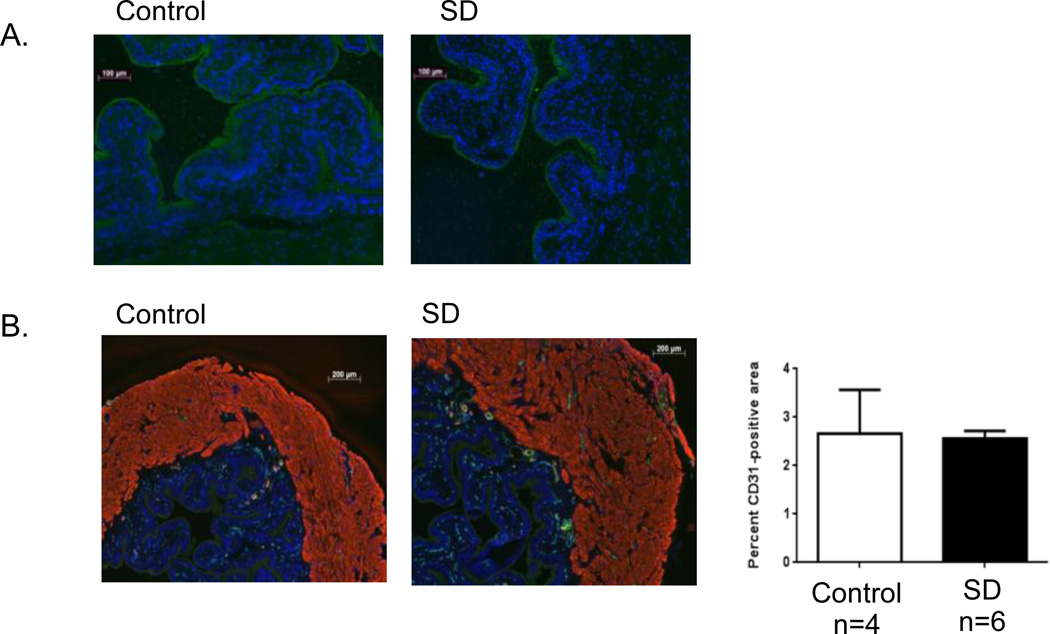
Figure 5.
Urothelial integrity and bladder vascularity are preserved in SD. Immunostaining of bladder sections from control and SD stressed mice for: (A) the urothelium-specific transmembrane protein uroplakin II (green), counterstained with DAPI (blue), at 100×, and (B) the endothelial marker CD31 (green), α smooth muscle actin (red) and DAPI (blue), at 50×. Following quantification with ImageJ no significant difference in the relative area of endothelial cell staining (CD31 positive) between groups was detected. Data are presented as the mean ± SEM.

