
FIGURE 3. Gr-1dimCD11b+ and Gr-1hiCD11b+ cells differ by nuclear morphology.
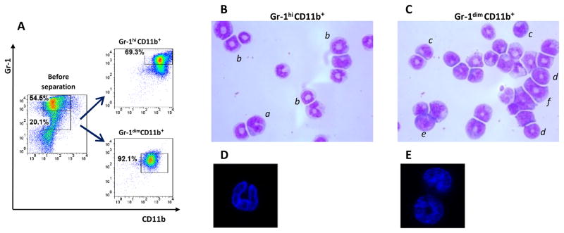
A, Enrichment of BMd24 cells for Gr-1dimCD11b+ and Gr-1hiCD11b+ populations. B, C, Nuclear morphology of Gr-1hiCD11b+ cells (B) and Gr-1dimCD11b+ cells (C) analyzed by light microscopy (cytospin preparations, Giemsa staining). a, PMN cell with segmented nucleus; b, PMN-like ring cell with ring-shaped nucleus and cytoplasmic center that is larger than the width of the ring; c, MNC-like cell with bean-shaped nucleus; d, MNC-like ring cell with a ring-shaped nucleus and a cytoplasmic center that is smaller than the width of the ring; e, MNC with closed nucleus; f, Precursor type of MNC-like ring cell showing a broad nuclear ring of round shape and regular contour and a small ring center. Original magnification ×1600. Cells are classified according to Biermann and coauthors (31). D, E Nuclear morphology of Gr-1hiCD11b+ cells (D) and Gr-1dimCD11b+ (E) cells analyzed by confocal microscopy (DAPI staining).