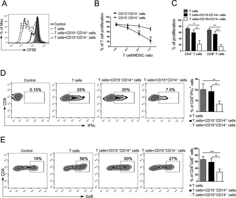
Figure 2. CD15HI MDSCs isolated from prostate cancer patients inhibit proliferation and activity of autologous T cells.
(A-C) CD15+CD14− granulocytic and CD15−CD14+ monocytic cell populations freshly enriched from metastatic prostate cancer patients’ PBMCs were cultured separately with 24 autologous T cells in presence of CD3-/ CD28-specific antibodies for stimulation. (A) Representative flow cytometry data showing T cell proliferation assessed by CFSE dilution after 3 days of co-culture. (B) Combined results of T cell proliferation assays from 5 patients showing percentage of total T cell proliferation at different T: myeloid cell ratios. (C) Proliferation of CD4+ and CD8+ T cells when incubated at 1:1 ratio with or without the indicated myeloid cell populations; means ± SD (n = 5). (D-E) CD15+CD14− myeloid cells inhibit production of IFNγ and granzyme B by activated CD8+ T cells. T cells were co-cultured with either one of myeloid cell populations at 1:1 ratio as above. The intracellular levels of IFNγ and granzyme B were measured using flow cytometry. Representative dot plots and bar graphs showing percentages of CD8+IFNγ+ T cells (D) and CD8+Granzyme-B+ T cells (E) after 3 days of culture; shown are means ± SD (n = 5). Statistically significant differences were indicated by asterisks.

