
Figure 5. The crystal structure of the chicken netrin-1/mouse DCC45 complex and the overlay of two complex structures.
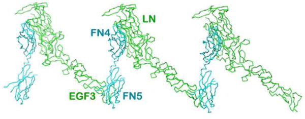
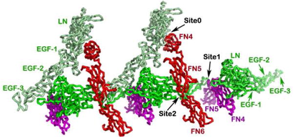
A. The netrin-1/DCC45 structure. The interactions between the DCC FN4 domain and the netrin-1 laminin-like domain along with the relative clustering seen in the crystal packing are highlighted. Note that the FN5/EGF-3 binding site is shared with what is seen in Fig. 2 at site 1. B. Overlay of the crystal structures of the chicken netrin-1/mouse DCC45 and the human netrin-1/human DCC56 highlights complementarity. All three binding sites are designated as binding site 0, binding site 1, and binding site 2. Netrin-1 molecules are colored in green and light green, respectively, whereas DCC receptors are colored in red and magenta, respectively.