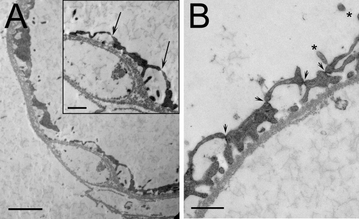
Fig. 2.
Electron micrographs of isolated inner medullary vas rectum (A) and type II descending thin limb of Henle's loop (B). A: vas rectum shows few fenestrations (arrows in inset), suggesting that it may be a descending vas rectum transitioning into an ascending vasa rectum. B: type II descending thin limb shows numerous tight junctions (arrowheads) and several microvilli (asterisks). Scale bars = 500 nm.

