Fig. 4.
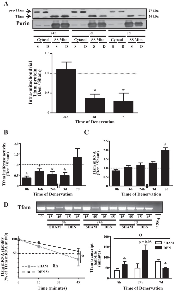
Denervation diminishes intramitochondrial localization of mitochondrial transcriptional factor A (Tfam) protein, represses activation of the Tfam promoter, and induces alterations in stability of the Tfam transcript. A: cytosolic and subsarcolemmal (SS) mitochondrial fractions were isolated from TA muscle denervated for 24 h, 3 days, and 7 days. Top: representative Western blot highlighting cytosolic form of Tfam (“pre-Tfam”) and cleaved mitochondrial Tfam; bottom: graphical representation of data from Western blot (n = 3–4 per time point). B: Tfam transcriptional activity in response to 8 h, 16 h, 24 h, 3 days, and 7 days of denervation, determined using a 1.1-kb proximal Tfam promoter-luciferase reporter construct in TA muscle (n = 4–7 per time point). C: Tfam mRNA content in response to denervation measured using quantitative PCR (n = 6 per group). D: degradation of Tfam mRNA in sham-operated or denervated TA muscle at 8 h, 24 h, or 7 days postsurgery. Top: representative ethidium bromide-stained agarose gel; bottom: graphical representation of rate of decay (left) used to calculate half-life of Tfam (right) at a given time point. A reaction in the absence of cDNA is shown as a negative control. Half-lives were calculated as described in materials and methods (n = 5 per time point). *P < 0.05 vs. sham at the same time point. αP < 0.05, main effect of treatment. Values are means ± SE.

