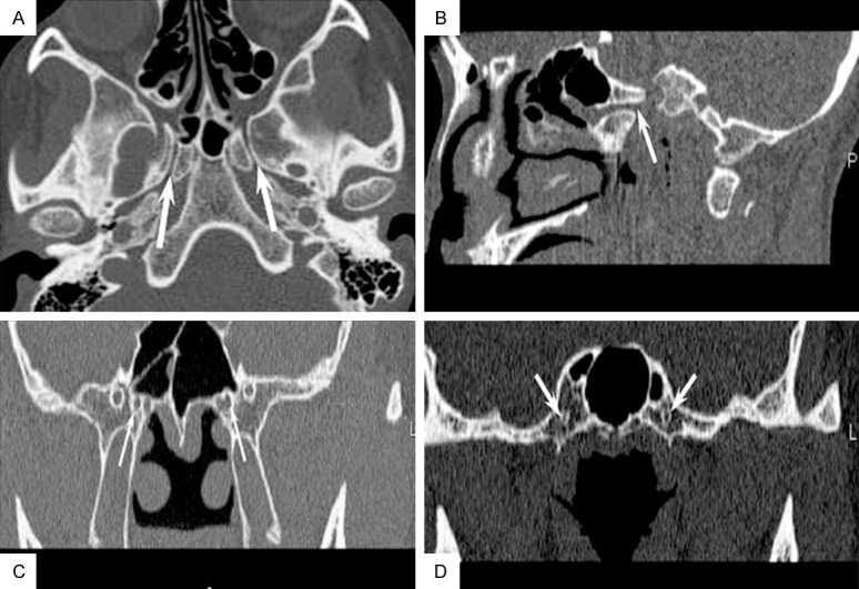
Figure 1.

On high resolution CT, axial-section (A) and oblique sagittal reconstruction (B) images show symmetric bilateral pterygoid canals (white arrows) with a narrow middle and relatively wider anterior and posterior openings connecting the pterygopalatine fossa and foramen lacerum shows; coronal section images detect oval-shaped anterior (C) and median openings (D) of pterygoid canals (white arrows).
