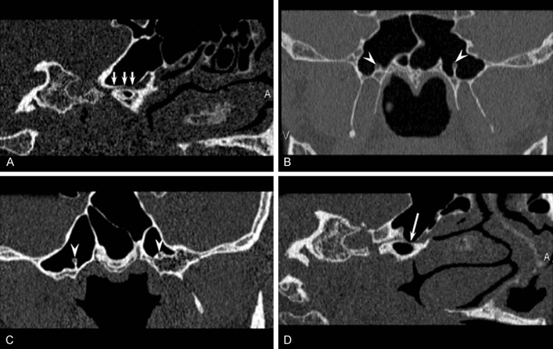
Figure 2.

Thin-section CT reconstruction images show bilateral pterygoid canals within the sphenoid sinus floor (white arrows) (A), inside the sphenoid sinus (arrowheads) (B), the right pterygoid canal inside the sphenoid sinus and the left pterygoid canal partially inside the sphenoid sinus (arrowheads) (C), and the superior wall of the pterygoid canal is missing (white arrow) (D).
