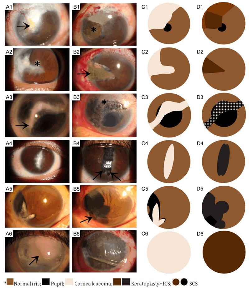
Figure 2.

A 27-year-old man whose left eye suffered from penetrating injury 19 years ago and has no light perception. A1, C1. A full-thickness corneal amyloidosis (arrow) with a rough surface before surgery. B1, D1. Partial LK combined with ICS (asterisk) was performed. 3 years later, the graft was smooth and flat, and the pigmentation was homogeneous. A 30-year-old man whose left eye was injured by a stick for 20 years ago and has light perception only. A2, C2. A full-thickness old scar in the 9 o’clock position with superior iridodialysis and traumatic white cataract (asterisk). B2, D2. Partial LK with ICS (arrow), cataract extraction and iridodialysis repair were performed simultaneously. A 34-year-old man whose left cornea was perforated by fireworks 20 years ago and has no light Perception. A3, C3. In the weak junction of the penetrating scar site and the irregular leucoma (arrow). B3, D3. only SCS was performed (asterisk). Disuse exotropia was also corrected. A 27-year-old man whose left cornea was perforated by a pair of scissors 21 years ago and has light perception only. A4, C4. Corneal leucoma, atretopsia and anterior synechia were diagnosed. B4, D4. To obtain good resistance to the tension at the scar junction and to prevent the dye from infiltrating into the eye, we separated the scar into two parts along the centre of the scar and used ICS (arrow), respectively. The two parts were connected but not cut through. A 46-year-old man whose right cornea was perforated by a broken glass10 years ago and has light perception only. A5, C5. Cornea leucoma, traumatic cataract and iridodialysis were diagnosed. B5, D5. Local LK (arrow), two-pocket ICS, cataract extraction, IO L implantation and coreoplasty were performed simultaneously. Visual acuity at the end of follow-up was 0.5. A 44-year-old man whose right eye was injured by fireworks 22 years ago and has no light perception. A6, C6. Whole cornea leucoma and central band keratopathy (arrow) were diagnosed. B6, D6. We performed calcified plaque scraping, EDTA chelation and a single whole corneal pocket ICS with no sutures. A therapeutic contact lens was worn for two weeks.
