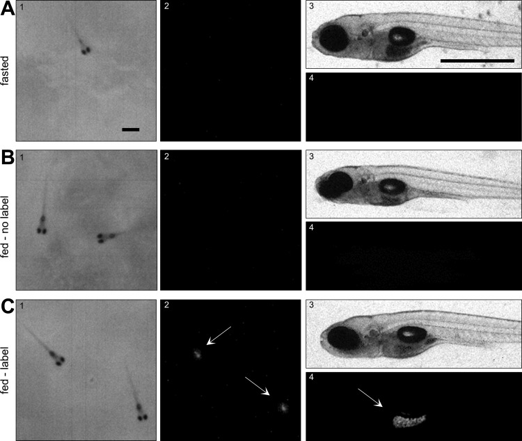
Fig. 3.
High-resolution images of the ingested DiR′ dye. Fasted zebrafish larvae (7 dpf) were exposed to none (A), nonlabeled (B), or labeled (C) paramecia for 30 min before euthanasia in iced water, agar embedding, and imaging with the infrared macroscope using the transmitted (1) or FL (2) light mode, and a Zeiss microscope using brightfield (3) and fluorescence (4) microscopy. Fish were euthanized to minimize digestion. Arrows indicate labeled paramecia in the larvae's intestine. Scale bar reflects 1 mm in both images.

