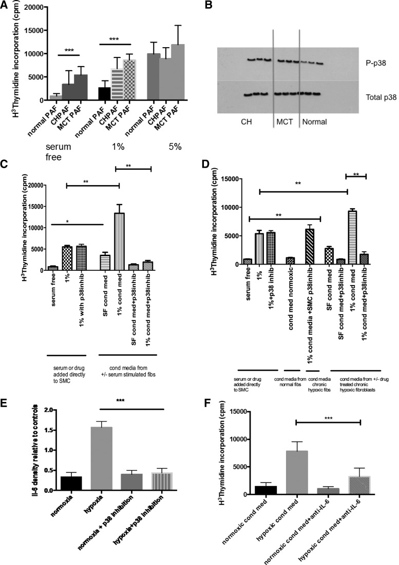
Fig. 1.
Proproliferative fibroblast phenotype and paracrine smooth muscle proliferation mediated by p38 MAPK in fibroblasts from experimental models of pulmonary hypertension (PH). A: cells were isolated from the 2nd order division of the pulmonary artery and grown in normoxic culture. The effect of serum stimulation was observed on these cells. At baseline with no serum stimulation, the chronic hypoxic (CH) and monocrotaline (MCT) pulmonary artery fibroblast (PAF) had an increased proliferative rate compared with the PAF derived from normoxic wild-type rats. This was also seen with low dose 1% serum stimulation. The effect was lost at 5% serum stimulation, which may reflect cell contact inhibition. The thymidine incorporation assay was used to assess the proliferation of the cells, and the results presented are representative of 3 experiments with triplicate values in each experiment. Values are means ± SE. ***P < 0.001. B: this immunoblot shows that under normoxic conditions the PAFs derived from both CH and MCT animals show a constitutive activation of p38 MAPK with increased levels of p-p38 detected. Cells were from passage 3 and were quiescesed in serum free medium for 24 h before harvest. Immunoblot for phospho-p38 and total p38 MAPK was performed and representative of 3 experiments. C: PAFs derived from MCT animals were isolated and then quiescesed for 24 h in serum free media. After 24 h, cells were stimulated with either 1% serum or some remained in serum free media ± SB203580 (p38 inhib). The conditioned media were aspirated from the wells after 24 h and passed through a cell sieve to give conditioned media from serum free cells ± SB203580 (SF cond med, SF cond med + p38 inhib) and 1% stimulated cells ± SB203580 (1% cond med, 1% cond med + p38 inhib). The conditioned media were added to smooth muscle cells for 48 h before a proliferation assay was performed. Both SF cond med and 1% cond med resulted in increased proliferation of pulmonary artery smooth muscle cells (PASMCs) while this effect was lost in the conditioned media from the PAFs, which had been coincubated with SB203580. There was no effect of SB203580 directly on the PASMCs. Values are means ± SE. Data shown from 3 experiments. **P < 0.005. D: PAFs derived from CH animals were isolated and then quiescesed for 24 h in serum free media. After 24 h, cells were stimulated with either 1% serum or some remained in serum free media ± SB203580 (p38 inhib). The conditioned media were aspirated from the wells after 24 h and passed through a cell sieve to give conditioned media from serum free cells ± SB203580 (SF cond med, SF cond med + p38 inhib) and 1% stimulated cells ± SB203580 (1% cond med, 1% cond med + p38 inhib). The conditioned media were added to normal PASMCs for 48 h before a proliferation assay was performed. Both SF cond med and 1% cond med resulted in increased proliferation of PASMCs while this effect was lost in the conditioned media from the PAF, which had been coincubated with SB203580. There was no effect of SB203580 directly on the PASMCs. Values are means ± SE. Data shown from 3 experiments. *P < 0.05; **P < 0.005. E: PAF from normal animals were exposed to 48 h normoxia or hypoxia ± SB203580. The supernatant was collected and analyzed using cytokine array. The above are representative of triplicate samples from 3 different animals. Exposure times for each blot was the same. The results for IL-6 are shown and are presented as a relative density plot vs. the positive control blots. ***P < 0.005. F: PASMCs are quiescesed and then incubated with conditioned media derived from normoxic or hypoxic PAF. The hypoxic conditioned media induced PASMC proliferation. When both the conditioned media and anti-IL-6 were added to the PASMCs, there was an inhibition in the proliferative stimulus. Results are means ± SE from 3 replicates from 3 different animals. ***P < 0.001.

