Fig. 7.
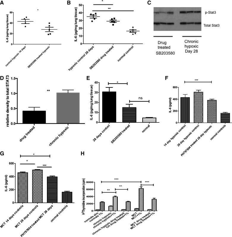
p38 MAPK inhibition in vivo leads to reduced IL-6 in experimental models of PH. A: lungs were isolated from CH animals in the prevention study with SB203580 and homogenized. The protein concentration was normalized by protein concentration as per BCA method. ELISA was used to analyze for IL-6 levels in the lung tissue. Data shown are means ± SE from triplicate samples from 4/5 animals in each group. B: lungs were isolated from normal and CH animals after treatment in reversal strategy with SB203580 and homogenized. The protein concentration was normalized by protein concentration as per BCA method. ELISA was used to analyze for IL-6 levels in the lung tissue. Drug-treated and normal control animals are at 28 days. Data shown are means ± SE from triplicate samples from 4/5 animals in each group. *P < 0.05 **P < 0.01. C and D: lungs from CH 28-day controls and SB203580-treated hypoxic animals were harvested and homogenized with a cocktail of phosphatase and kinase inhibitors. The protein concentration was quantified using BCA method. Equal concentrations were then loaded on a gel and blotted for phospho-STAT3 and total STAT3. Immunoblot shown is best representative of 3 experiments using lungs from 3 different animals with each condition. Densitometry is shown of other blots. **P < 0.001. E: lungs were isolated from MCT and normal control animals and homogenized. Inhibitor used was SB203580. The protein concentration was normalized by protein concentration as per BCA method. ELISA was used to analyze for IL-6 levels in the lung tissue. Data shown are means ± SE from triplicate samples from 4/5 animals in each group. *P < 0.05; ns is not significant by ANOVA. F: PH-797804 reduces serum IL-6 in reversal of CH-induced PH. Serum was collected from animals at the time of cardiac puncture and stored at −80°C until analysis could be performed. ELISA for IL-6 was performed on serum samples. Values shown are means ± SE. Samples were analyzed in duplicate and total animal number n = 11. ***P < 0.005. G: PH-797804 reduces serum IL-6 in reversal of MCT-induced PH. Serum was collected from animals at the time of cardiac puncture and stored at −80°C until analysis could be performed. ELISA for IL-6 was performed on serum samples. Values shown are means ± SE. Samples were analyzed in duplicate and total animal number n = 12. *P < 0.01; ***P < 0.005. H: fibroblasts undergo phenotypic switch back to normal after p38 MAPK inhibition. PAF were cultured from pulmonary arteries derived from normal, experimental models of PH (CH and MCT) and from animals after treatment with p38 MAPK inhibition PH-787904 for 2 wk. Cells were challenged with or without serum to assess proliferation; n = 3–4 per group. Experiment repeated 3 times. **P < 0.01, ***P < 0.001.

