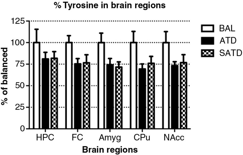
Fig. 5.
Levels of tyrosine in the different brain regions of the mouse after formula administration. Data are represented as mean±S.E.M. Groups of 7–8 mice received either a control condition (BAL), acute tryptophan depletion (ATD), or simplified acute tryptophan depletion (SATD) mixtures. HPC: hippocampus; FC: frontal cortex; Amyg: amygdala; CPu: caudate putamen; NAcc: nucleus accumbens.

