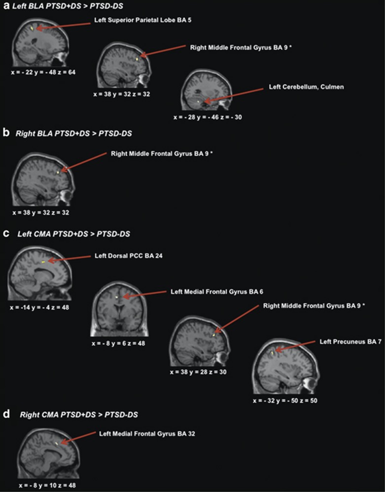
Figure 1.
(a) Brain areas representing greater connectivity to the left basolateral amygdala within PTSD+DS as compared with PTSD−DS; (b) brain areas representing greater connectivity to the right basolateral amygdala within the PTSD+DS as compared with PTSD−DS; (c) brain areas representing greater connectivity to the left centromedial amygdala within PTSD+DS, as compared with PTSD−DS; (d) brain areas representing greater connectivity to the right centromedial amygdala within PTSD+DS as compared with PTSD−DS. Statistical threshold p<0.005 uncorrected, k=10 for all two-sample t-tests. BLA, basolateral amygdala; CMA, centromedial amygdala; PCC, posterior cingulate cortex; PTSD+DS, dissociative subtype posttraumatic stress disorder group; PTSD−DS, non-dissociative posttraumatic stress disorder group. *Indicates the a-priori region-of-interest analysis.

