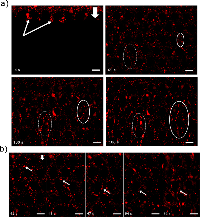
Figure 3.
(a) Rapid streamer formation in a short time scale (a few seconds). The scale bars are 50 μm. See accompanying video. The images were taken approximately at the middle of the channel height (z = 25 μm). In the top-left image, the arrows demarcate the advancing fluid meniscus. The ellipses demarcate two regions where streamers form. (b) An arrow demarcates a floc, which is first advected through the channel and then is attached to a micropillar wall at t = 47 s and finally at 95 s a streamer is formed. Scale-bars are 50 μm.

