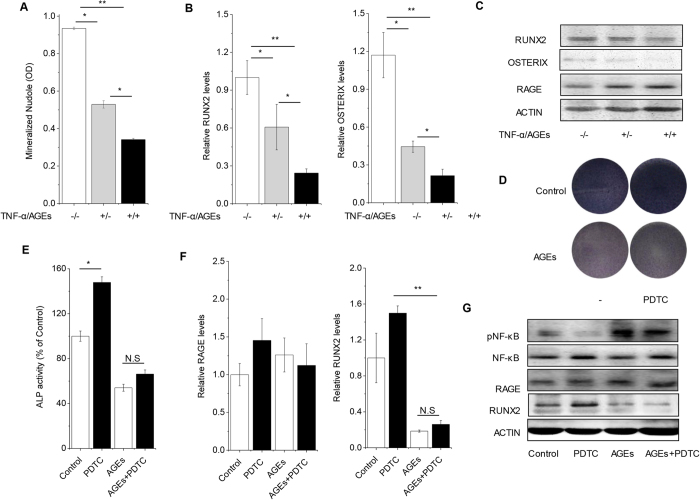
Figure 3. Effects of AGEs on osteogenesis of PDLSC in vitro.
(A) PDLSCs were treated with TNF-α and AGEs, which mimics the PDLSCs from periodontitis with diabetes mellitus persons. Quantification of Alizarin Red staining was examined when PDLSCs with TNF-α and AGEs were induced in osteogenic differentiation for 28 days (n = 3). (B) The expression of osteoblastic gene RUNX2 and OSTERIX was examined by Real-time PCR on day 7 (n = 3). (C) The expression of RAGE and osteoblastic gene were examined by western blot analysis on day 7. (D,E) PDLSCs were treated with AGEs and NF-κB inhibitor PDTC. The ALP activity staining were quantified on day 7 (n = 4). (F) The expression of RAGE and osteoblastic gene were examined by Real-time PCR on day 7 (n = 3). (G) The activation of NF-κB (phosphorylated p65, pNF-κB), RUNX2 and RAGE was examined by western blot analysis. Data (±SD) are representative of two (E) or three (A,B,F) independent experiments. Student’s t test was performed to determine statistical significance (*p < 0.05, **p < 0.01).

