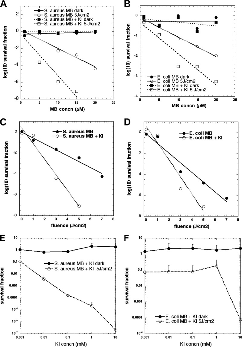
FIG 1.
Effect of KI on MB-mediated PDT of bacteria. S. aureus (A) or E. coli (B) bacteria (108 cells/ml) were incubated with a range of MB concentrations for 15 min followed or not by addition of KI and illumination with 5 J/cm2 of a 660-nm light. S. aureus (C) or E. coli (D) bacteria (108 cells/ml) were incubated with an MB concentration of 10 μM for 15 min followed by addition of KI (10 mM) and illumination with different fluences (0 to 7 J/cm2) of 660-nm light. (E and F) Different concentrations of KI (0.01 to 10 mM) were tested with MB-PDT against bacteria. (A to D) Best linear fit curves. S. aureus (E) or E. coli (F) bacteria (108 cells/ml) were incubated with 10 μM MB followed by addition of a range of KI concentrations and illumination with 5 J/cm2 of 660-nm light.

