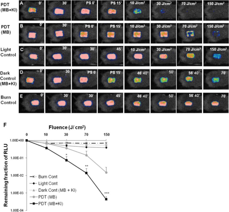
FIG 7.
(A to E) Successive bacterial bioluminescence images of representative mouse burns infected with 108 CFU of luminescent MRSA (USA300) and treated with PDT using a mixture of MB (50 μM) and KI (10 mM) (A) or PDT using MB (50 μM) at 30 min after bacterial inoculation and 15 min from PS application (B). PDT was carried out with a combination of 50 μl of a mixture containing MB and KI or MB alone and 150 J/cm2 red light (660 ± 15 nm; 100 mW/cm2). (C) Light alone. (D) Application of a mixture of MB and KI but without red light illumination (dark control). (E) Burn control without any treatment. (F) Dose response of mean bacterial bioluminescence of mouse burns infected with MRSA (USA300) after treatment with light alone, a mixture of MB (50 μM) and KI (10 mM) (dark control), PDT using MB (50 μM) alone, or a mixture of MB (50 μM) and KI (10 mM).

