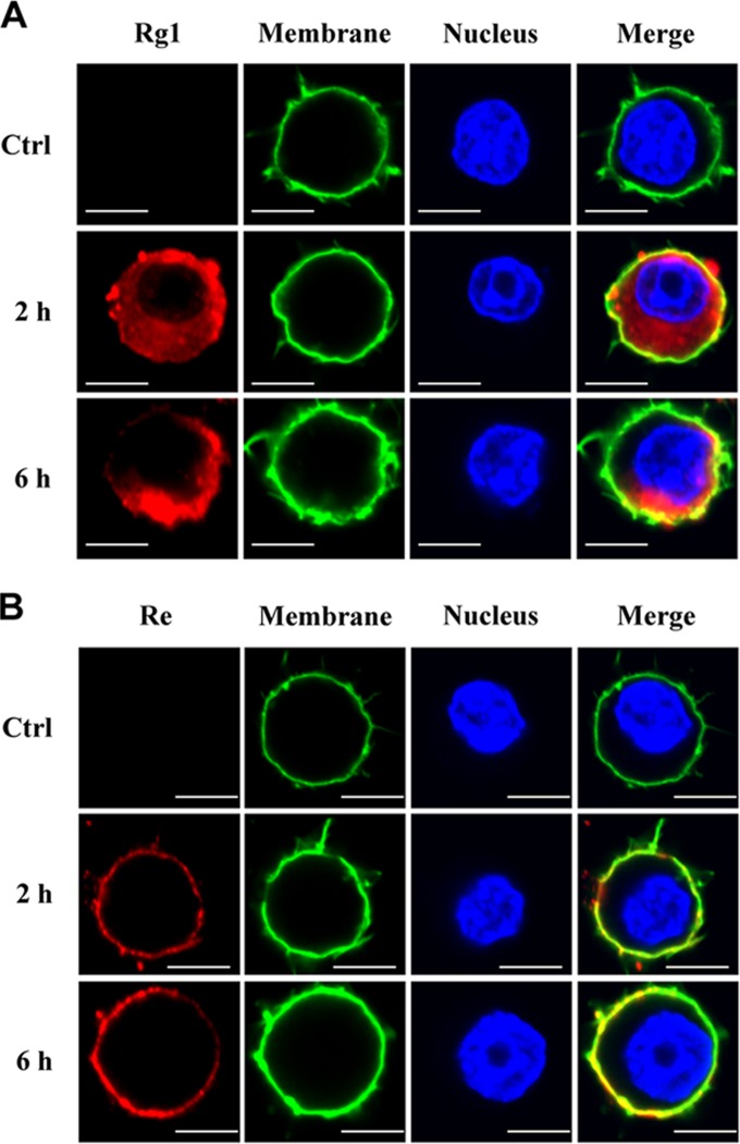
FIG 3.
Localization of Rg1 and Re in macrophages. (A) Rg1 was localized both inside and outside cells. RAW264.7 cells were incubated with Rg1 (50 μg/ml) for 2 or 6 h and then stained with primary antibody and Alexa Fluor 594-labeled secondary antibody (red). Cells without Rg1 treatment were set as the control (Ctrl). Images are representative of three independent experiments. Bars, 10 μm. (B) Re was localized only on the cell membrane. RAW264.7 cells were treated in the same way as described for panel A except that Re and anti-Re antibody were used instead of Rg1 and anti-Rg1 antibody. Bars, 10 μm.

