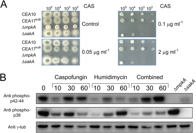
FIG 3.
Sensitivity of the ΔsakA and ΔmpkA strains to different CAS concentrations and Western blot analysis. (A) The indicated numbers of conidia were spotted on AMM agar plates with or without different concentrations of CAS. (B) The wild-type strain was stressed by humidimycin and CAS alone or in combination, and samples were collected at the indicated time points (minutes after stress). The anti-phospho p42-44 antibody was used to detect the phosphorylated form of MpkA, while the anti-phospho p38 antibody was used for SakA phosphorylated forms. As negative controls, proteins extracted from the ΔsakA and the ΔmpkA strains under nonstressed condition were used. γ-Tubulin was used as a control (Anti γ-tub).

