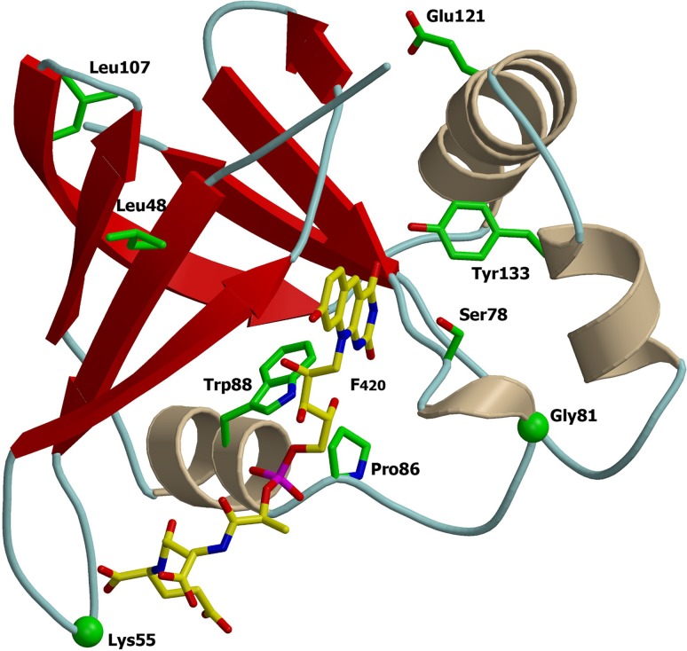
FIG 3.
Ribbon representations of Mycobacterium protein Ddn (PDB code 3R5R). Mutated residues identified are represented on the three-dimensional (3D) protein structures. The F420 is depicted with carbon atoms in yellow. The images were obtained using a consecutive combination of the MolScript (37) and Raster3D (38) programs.

