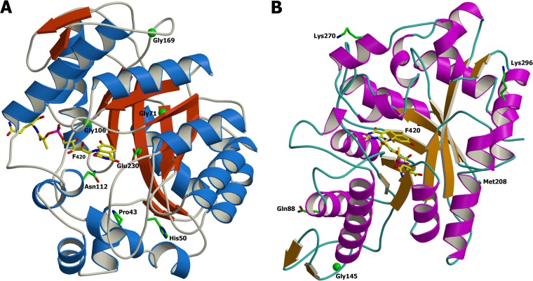
FIG 4.
Ribbon representation of the crystal structure of Mycobacterium FGD1 (PDB code 3BY4) (A) Mutated residues identified are represented on the 3D protein structures. The F420 is depicted with carbon atoms in yellow. Phylogenetic amino acid substitutions reported by Feuerriegel et al. (35) are shown (B). These residues are found on the protein surface. The image was produced consecutively using the MolScript (37) and Raster3D (38) programs.

