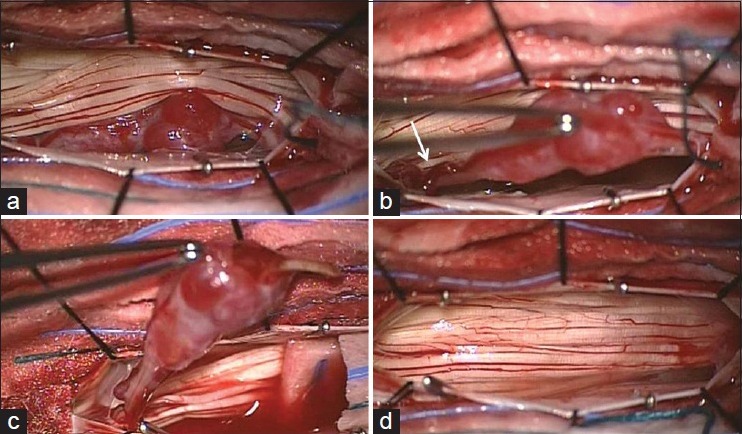
Figure 2.

Intraoperative view of lesion. The lesion appeared as a white reddish elliptical-nodular lesion intimately involved with the nerve root displacing the cauda equina (a) Enlarged vessels were also evident along the nerve root (b, white arrow). After dissecting the lesion from cauda equina and cutting the originating nerve root distally (c) the lesion was totally removed (d)
