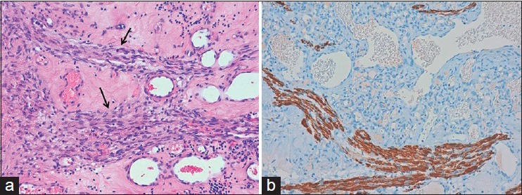
Figure 3.

Histological picture (a) of the surgical specimen shows many, variably sized, thin-walled vascular channels lined by endothelial cells with interposed nerve fibers (black arrows) (H and E, original magnification ×20). Immunohistochemical staining (b) for neurofilament protein highlights the nerve fibers interspersed among the vascular spaces. The lesion was also positive at S-100 staining (not showed) (original magnification ×20)
