Fig. 4.
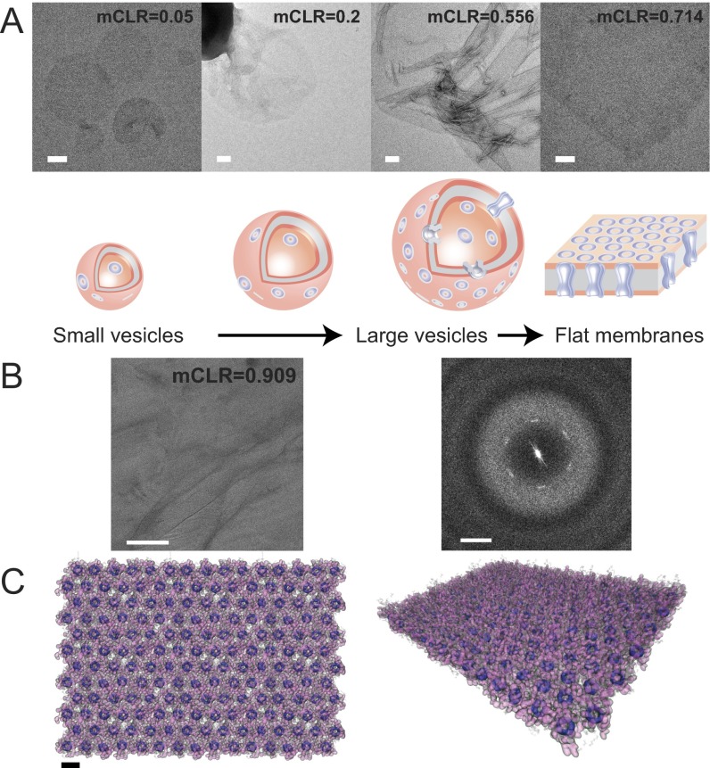
The concentration of PAP channels influences the morphology of self-assembled channel–lipid aggregates. (A) Negative-stain EM images show the representative morphologies of aggregates formed at different molar channel-to-lipid ratios (mCLRs). Increasing mCLRs from 0.05 to 0.2 to 0.625 to 0.714 resulted in morphology transitions from small vesicles to large vesicles and finally to flat membranes. Scale bars are 100 nm. (B) (Left) A cryo-EM image of a self-assembled channel–lipid aggregate formed at an mCLR of 0.909 that was frozen in trehalose. (Scale bar, 100 nm.) (Right) The power spectrum, which reveals blurry first-order diffraction spots, indicating that the PAP channels form a somewhat ordered hexagonal array with lattice dimensions of a = b = 21 Å and γ = 120°. (Scale bar, 0.5 nm−1.) (C) Top (Left) and tilted (Right) views of a molecular model of a partially ordered array of hexagonally packed PAP channels in a lipid bilayer. The inner channel rings (dimethoxy benzene) are rendered as opaque dark violet surfaces, phenylalanine arms are rendered as translucent purple surfaces, and lipid molecules are rendered as transparent gray polygons. For clarity, water is not shown. The model is the result of an MD simulation guided by the ordering observed in the cryo-EM images. (Scale bar, 2.1 nm.)

