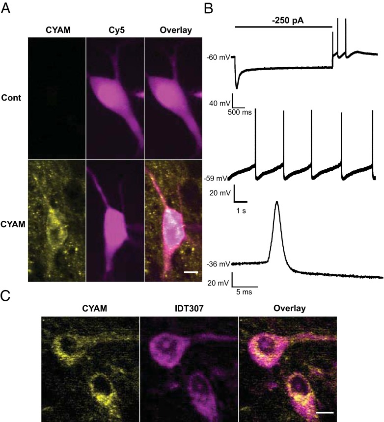
Fig. 4.
CYAM in SNc DA neurons. (A, B) Electrophysiological confirmation of SN DA neuron CYAM accumulation. During whole-cell patch-clamp recordings, cells from control and CYAM-treated slices were filled with patch-pipette solution containing 10 μM sulfo-Cy5-COOH (Cy5). (A) Pseudocolored summed z-projections of CYAM (yellow) and Cy5 (magenta) fluorescence signals. (Scale bar: 10 μm.) (B) Representative current-clamp voltage traces of electrophysiological characteristics used to establish dopaminergic identity of a cell. (Top) Characteristic Ih voltage sag and spontaneous rebound spiking activity of a neuron that was current-clamped at −60 mV, followed by a 4-s, −250-pA current injection. Slow pacemaker activity (Middle) and broad action potentials (Bottom) are characteristic of SNc DA neurons. (C) Colocalization of CYAM (yellow) and two monoamine neurons in the SNc identified with IDT307 (magenta). (Scale bar: 10 μm.)

