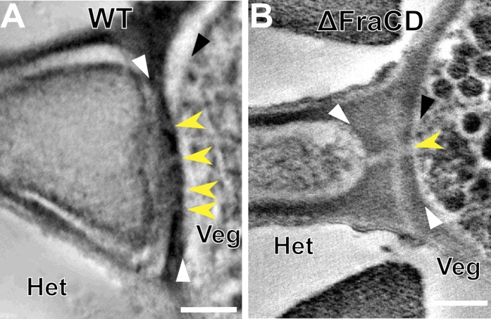
Fig. 2.
Heterocyst-vegetative cell septa in WT and fragmentation mutants. (A) Electron tomographic image of a WT heterocyst junction. White arrowheads point to the edges of the septum. Yellow arrowheads show the channels that connect the heterocyst and the vegetative cell. (B) Electron tomographic image of the CSVT22 (ΔfraCD) heterocyst junction. The septum in this mutant is thicker and only 1–2 channels are present compared with WT. The yellow arrow points to the only channel observed in this tomogram. Black arrowheads point to the plasma membrane in vegetative cells in each panel. All tomographic images are composed of 10 superimposed 2.2-nm tomographic slices. Het, heterocyst; Veg, vegetative cell. (Scale bar: 200 nm.)

