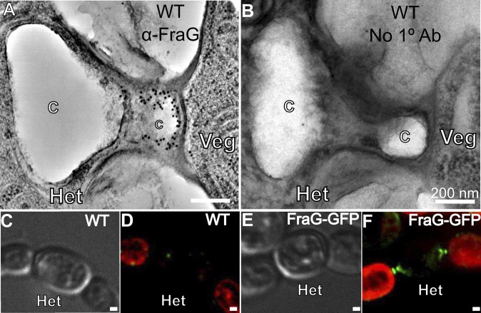
Fig. 4.
Subcellular localization of FraG in Anabaena heterocysts. (A) Immunogold labeling of WT Anabaena using antibodies (black dots) raised against the N-terminal coiled-coil domain of FraG. (B) Control immunogold labeling of WT using only secondary antibody; no dots. (C) Light transmission micrograph of WT Anabaena grown under N− conditions. (D) Autofluorescence of the same cells shown in C. Heterocysts do not show autofluorescence due to loss of PS II chlorophyll. (E) Light transmission micrograph of the CSAM137 mutant (FraG-GFP) grown under N− conditions. (F) Autofluorescence (red) and GFP fluorescence (green) of the same cells shown in E. GFP fluorescence locates FraG at the poles of the heterocysts. C, Cyanophycin; Het, heterocyst; Veg, vegetative cell. (Scale bar: 200 nm.)

