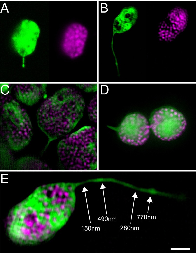
Fig. 4.
Examples of fluorescent stromules in N. benthamiana chloroplasts visualized by 3D structured illumination microscopy (3D-SIM). (A and B) Comparison of confocal laser scanning microscopy (A; also shown in Fig. S11) and 3D-SIM (B; also shown in E and in Movie S3) to visualize chloroplast structure (Left: stromal GFP, green; Right: thylakoid chlorophyll, magenta). In particular, note the improved resolution of stromule width and the clarity of the thylakoid grana in the 3D-SIM z-slice (B). (C) 3D-SIM z-slice image of mesophyll chloroplasts with stromules. (D) An epidermal chloroplast connected by a thin bridge that contains both stroma and thylakoids also has a stromule (Left), as shown by SIM. (E) 3D-SIM reveals variability in stromule width. Stromal GFP, green; chlorophyll autofluorescence, magenta. (Scale bar: 2 μm.) One z-slice from a 3D-SIM reconstruction is shown, with measured stromule diameters labeled at indicated positions (white arrows).

