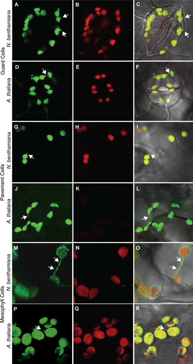
Fig. S3.
Representative images of chloroplasts and leucoplasts in N. benthamiana and A. thaliana visualized with confocal laser scanning microscopy. Guard cells (A–C and D–F), pavement cells (G–I and J–L), and mesophyll cells (M–O and P–R) all contain chloroplasts in N. benthamiana, as do A. thaliana guard cells (E) and mesophyll cells (Q); however, A. thaliana pavement cells (J–L) have leucoplasts that lack chlorophyll (K). Green, stromal GFP; red, chlorophyll autofluorescence; gray, transmitted light. Some stromules are indicated by white arrows. Error bars indicate SE.

