Fig. 1.
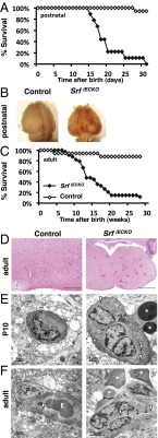
Premature death and cerebral hemorrhages in SrfiECKO animals. (A) Kaplan–Meier plot (percent survival) of control (n = 16) and SrfiECKO (n = 9) animals upon postnatal Srf deletion. (B) Brains revealing hemorrhages in SrfiECKO mice (P17). (C) Kaplan–Meier plot (percent survival) of control (n = 16) and SrfiECKO (n = 33) animals upon adult Srf deletion. (D) H&E staining of adult control and SrfiECKO brain sections, revealing multiple hemorrhages in SrfiECKO brains (representative acute-stage animal shown). [Scale bars, 200 μm (Left) and 1,000 μm (Right).] (E) Electron microscopic images highlight intact blood vessels in P10 control animals and hemorrhages in SrfiECKO brains. (Scale bars, 1 μm.) (F) Electron microscopic images highlight intact blood vessels in adult control animals and hemorrhages in SrfiECKO brains. (Scale bars, 2 μm.) Arrows highlight obliterated vessel lumen; arrowheads point to tight junctions; white asterisks mark extravasated erythrocytes. E, intraluminal erythrocyte; P, pericyte.

