Fig. 2.
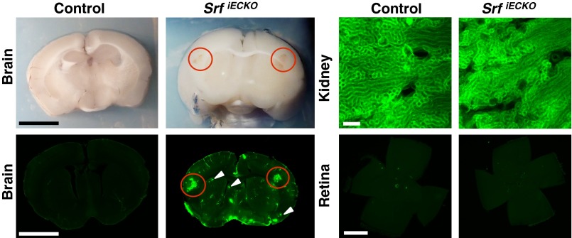
Extravasation at localized sites of hemorrhage. Sulfo-NHS-LC-Biotin extravasations at localized sites of hemorrhage in SrfiECKO, but not control brains, as visualized by blood coloring in brightfield (Upper Left) and fluorescence imaging (Lower Left) of an identical brain section. (Scale bars, 1 cm.) Red circles indicate larger-volume extravasations; white arrowheads indicate microbleeds. (Upper Right) Kidneys, composed of fenestrated endothelium, display constitutive dye extravasation. (Scale bar, 200 μm.) (Lower Right) Retinal flat-mounts evidence lack of extravasation in the retina (n = 4). (Scale bar, 1,000 μm.)

