Fig. 4.
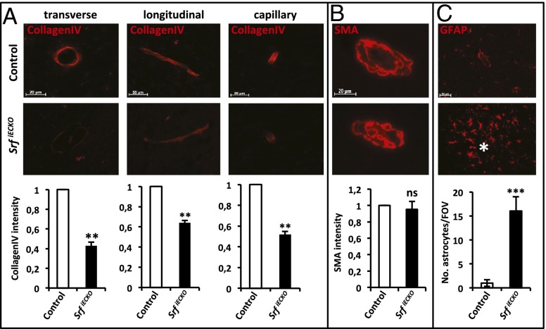
SrfiECKO cerebral microvessels show reduced collagen IV staining and elevated astrocyte recruitment. (A) Collagen IV staining of P10 cerebral microvessels in control (Upper) and SrfiECKO (Middle) brains, as quantified by immunofluorescence signal intensity (Lower). (Left) Transverse section. (Scale bar, 20 μm.) (Center) Longitudinal section. (Scale bar, 50 μm.) (Right) Capillary. (Scale bar, 20 μm.) (B) Smooth muscle actin (SMA) staining of adult control (Upper) and SrfiECKO (Middle) brains revealed no significant difference in smooth muscle cell presence (Lower). (Scale bar, 20 μm.) (C) Astrocytes stained by glial fibrillary acidic protein (GFAP) accumulate specifically around lesion sites in adult SrfiECKO brains (Middle; hemorrhage highlighted by asterisk). Quantification of astrocyte number per field of view (n = 5; Lower). (Scale bar, 50 μm.) Data are presented as means ± SEM. **P < 0.01; ***P < 0.001; ns, not significant.

