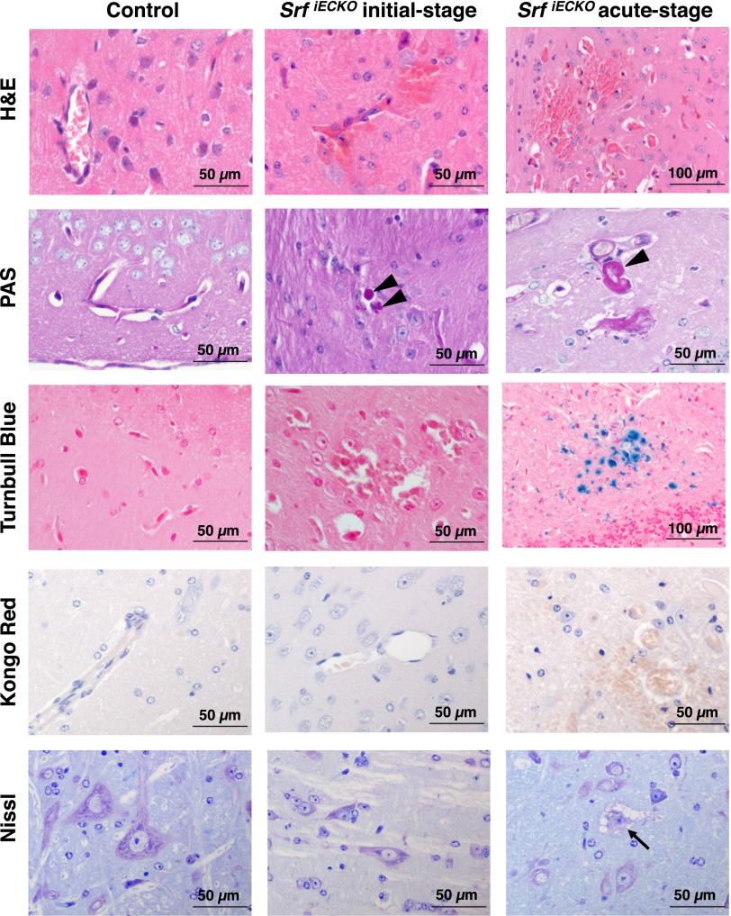
Fig. S5.
Adult SrfiECKO animals display common and differential pathological characteristics at initial vs. acute stages. Adult control (Left), initial-stage SrfiECKO (Center), and acute-stage SrfiECKO (Right) animals are compared regarding disease-associated processes. H&E staining reveals brain hemorrhages in initial-and acute-stage SrfiECKO animals, with erythrocytes being localized in the surrounding tissue outside blood vessels. PAS reactivity (arrowheads) shows plasma extravasation in both SrfiECKO disease stages. Turnbull Blue staining, indicating aged iron deposits, is seen in acute-stage SrfiECKO brains only. Kongo Red, a marker for amyloid plaques, which occur in cerebral amyloid angiopathy patients, is negative in control and both SrfiECKO stages. Nissl staining indicates neuronal degeneration only in the acute-stage SrfiECKO animals (arrow). (Scale bars, 50 and 100 μm, as indicated in the images.)

