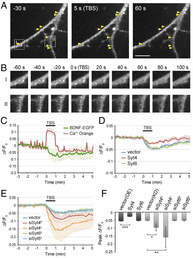
Fig. 6.
Syt4, but not Syt6, regulates activity-induced secretion of BDNF-EGFP in dendrites. (A) Representative images of hippocampal neurons transfected with control vector and BDNF-EGFP, taken at 30 s before, and 3 s and 60 s after TBS application. Arrows, BDNF-EGFP puncta showing fluorescence reduction after TBS. (Scale bar, 10 μm.) (B) Boxed areas I and II in A are shown at a higher resolution. (Magnification, 1.12 × 1.12.) (C) A representative trace of average BDNF-EGFP and Ca2+ Orange fluorescence changes at dendrites in control neurons (shown in A and B), with TBS period marked by the bar. Fluorescence levels were normalized by that before TBS (n = 10). Error bars, SD. (D) Traces of average BDNF-EGFP puncta fluorescence in dendrites of control and Syt4- and Syt6-overexpressing neurons (n = 4 cultures in each group). Error bars, SEM. (E) Traces of average BDNF-EGFP puncta fluorescence in dendrites of control and siSyt4- or siSyt6-transfected neurons (n = 3–5 cultures each). Error bars, SEM. (F) Average peak reduction of BDNF-EGFP puncta fluorescence in dendrites induced by TBS, normalized to the fluorescence intensity before TBS. Error bars, SEM (n = 3–5 cultures each; *P < 0.01, **P < 0.005, ***P < 0.001 by one-way ANOVA and Tukey post hoc test).

