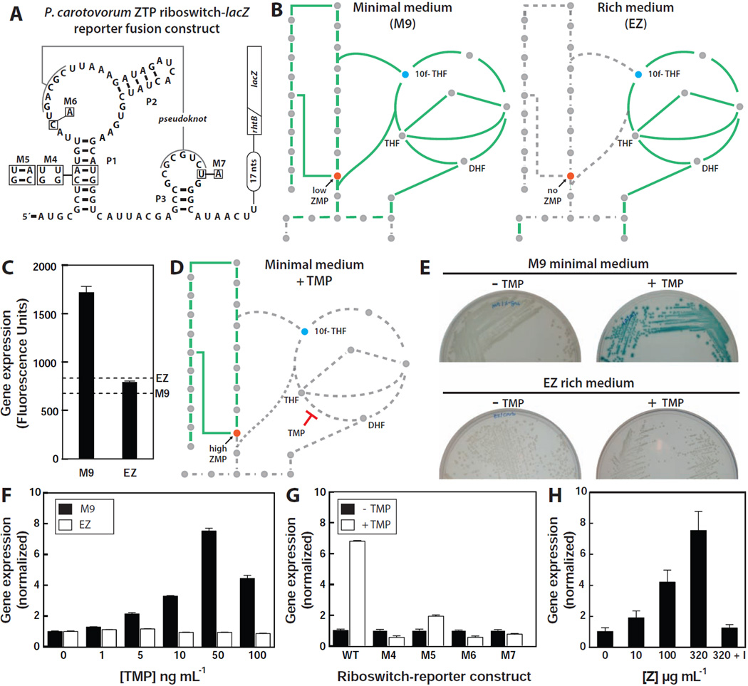
Figure 6. ZMP/ZTP-induced Reporter Gene Expression Controlled by a Riboswitch Associated with Pectobacterium carotovorum rhtB.
(A) Sequence and secondary structure of the WT ZTP riboswitch associated with the rhtB gene of P. carotovorum and various mutants fused to a β-galactosidase reporter gene (lacZ). The diagonal line indicates the fusion of the first eight codons of rhtB to the ninth codon of lacZ. Other annotations are as described for Figure 3A.
(B) Schematic representation of the expected metabolic flux in minimal (left) and rich (right) medium. Solid green lines indicate pathways with flux whereas dashed gray line represents pathway with low or no flux.
(C) Plot of reporter gene activity for E. coli cells containing the riboswitch reporter construct and grown on minimal (M9) or rich (EZ) medium supplemented with X-gal (See Extended Experimental Procedures). Dashed lines represent the background measurements for M9 and EZ media in the absence of cells.
(D) Schematic representation of the expected metabolic flux in minimal medium containing trimethoprim (TMP). Other annotations are as described for B.
(E) Agar plate assays with E. coli cells containing the riboswitch reporter construct and grown under the conditions as indicated in media supplemented with X-gal.
(F) Plot of the relative reporter gene expression levels with increasing concentrations of TMP relative to no treatment either in minimal (M9) or rich (EZ) medium.
(G) Plot of the relative β-galactosidase activity in cells containing the WT or mutant riboswitch reporter constructs depicted in A in the absence or presence of 0.1 µg mL−1 TMP, as indicated, in minimal medium.
(H) Plot of the relative β-galactosidase activity of cells carrying the WT riboswitch reporter construct upon the addition of increasing concentrations of Z riboside in rich medium. “I” indicates addition of 1 mM 5´-amino-5´-deoxyadenosine, a selective inhibitor of adenosine kinase.

