
Figure 1.
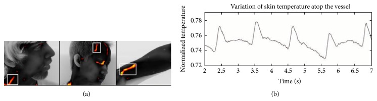
Pulse computation from thermal imaging data. (a) Collection point on the carotid arteriovenous complex, the frontotemporal region, and the wrist of the subject. (b) Temperature profile after removing frequency signals lower than 0.67 Hz (40 bmp) and higher than 1.67 Hz (100 bmp) (adapted from [9]).