
Figure 2.
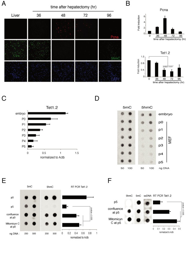
Regulation of Tet1 in mouse proliferating cells. (A) Immunofluorescence analysis of Pcna and 5hmC in liver after hepatectomyzed the indicated time. DAPI is used for nuclei staining. (B) RT-qPCR of Tet1 and Pcna mRNA in liver after hepatectomy. (C) RT-qPCR of Tet1 mRNA in mouse embryo or in MEFs after several passages. (D) Dot-blot analysis of 5hmC and 5mC in mouse embryo or in MEFs after several passages. (E) Left panel: dot-blot analysis of 5hmC and 5mC in MEFs just extracted (p0) or maintained proliferating (p5) or blocked (by confluence or Mitomicyn C) from p0. Right panel: RT-qPCR of Tet1 mRNA in MEF in the same conditions. (F) Same experiment as in (E), but the blocking of proliferation was induced at p5 and maintained for other 5 days. Error bars represent the standard deviation of three independent experiments. P-value was calculated by using t-test.