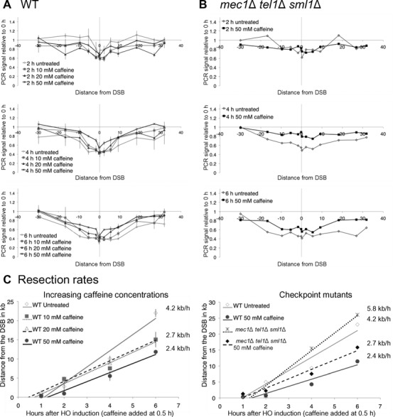
Figure 2.

Caffeine treatment impairs resection independent of Mec1 and Tel1. Cells were arrested in G2/M by nocodazole treatment 3 h prior to HO induction. (A) Resection in strain JKM179 without caffeine treatment (diamonds), or when 10 mM caffeine (squares), 20 mM caffeine (triangles) or 50 mM caffeine (crosses) was added 0.5 h after HO induction. Error bars represent ranges. (B) Resection in JKM179 mec1Δ tel1Δ sml1Δ without caffeine treatment (gray) and when 50 mM caffeine was added 0.5 h after HO induction (black) at different times after HO induction. (C) Rates were calculated by determining the time and distance when the PCR signal fell to 75% of signal at 0 h (36). These values were plotted on time-versus-distance graphs, and the rates were determined by linear regression analysis. Left graph represents resection measurements seen in (A) error bars represent the range. Right graph represents resection measurements seen in (B).
