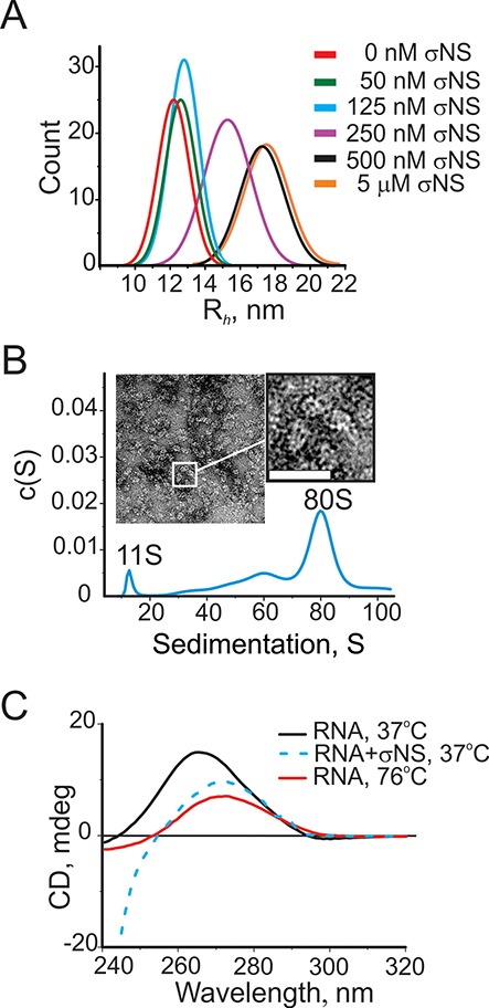
Figure 4.

Assembly of σNS ribonucleoproteins with long ssRNAs. (A) Size (Rh) distributions of a 3.6 kb-long ssRNA alone (1 nM, red), and upon addition of σNS, measured by FCS. (B) Sedimentation velocity of the long ssRNA (25 nM), incubated with σNS (10 μM). A large 80S complex with fewer smaller species is formed. Inset – negative stain EM micrograph of the σNS, bound to long ssRNAs, revealing multiple spherical ribonucleoproteins. Bar = 25 nm. (C) Circular dichroism (CD) spectra of the long ssRNA (as in panels A and B), reveal RNA secondary structure destabilization upon incubation with the ARV σNS. CD spectra for folded RNA alone at 37°C (15 nM, black) and thermally unfolded RNA at 76°C (red) are shown along with the CD spectrum of the RNA, incubated with σNS for 5 min (15 μM, blue dashed line). Note the significantly lower intensity of the 263 nm peak in the latter, characteristic for an A-form double-stranded helix, with its shift toward 271 nm, typical for single-stranded RNA. Below 240 nm, the CD signal from RNA is mainly dominated by the contribution of the protein.
