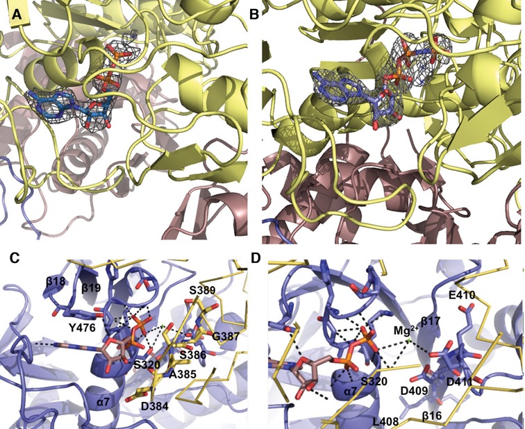
Figure 3.

Nucleotide binding by MRB1590. (A) Fo − Fc omit map (blue mesh, contoured at 3 σ and calculated to 2.6 Å) in which the ADP was omitted from the refinement. Clear electron density for the ADP molecule bound near the P-loop region is observed. (B) Fo −Fc omit map (blue mesh, contoured at 3σ to 3.0 Å) showing density for the bound AMP–PNP, which was omitted from the refinement. (C) ADP binding by MRB1590. One subunit of the MRB1590 dimer is colored blue and the other is yellow. The specific amino acids in the Walker A or P-loop are highlighted as sticks. Also shown is the tyrosine residue, Tyr476, which stacks with the adenine base. The other subunit in the dimer, colored yellow, contributes the signature region, 385–389. The residues in this region are highlighted as yellow sticks and labeled. (D) Close up of the magnesium coordination site. The residues responsible for magnesium binding come from the Walker B motif, residues 407-411, which are located on β16. The magnesium ion is shown as a green cross and labeled.
