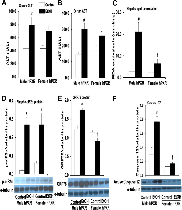
Fig. 5.
Markers of hepatotoxicity in control and binge EtOH-treated male and female hPXR mice. Male and female hPXR mice were orally administered saline (control; □) or EtOH (4.5 g/kg; ▪) every 12 hours for a total of three doses and were killed 4 hours after the final dose. Serum ALT (A) and AST (B) levels were determined as described in Materials and Methods. The extent of lipid peroxidation in liver tissues was quantified by measuring the thiobarbituric acid–reactive product, MDA (C). Western blots of liver homogenate (40 μg/lane) were probed with antibodies to phospho-elF2α (D), GRP78 (E), and caspase 12 (F). Bands were quantified and normalized to α-tubulin. Data represent the mean ± S.E.M. of two independent experiments from 3–4 mice/group. #P < 0.05 between male and female mice treated with saline (control) and EtOH; †P < 0.05 between male and female mice fed EtOH.

