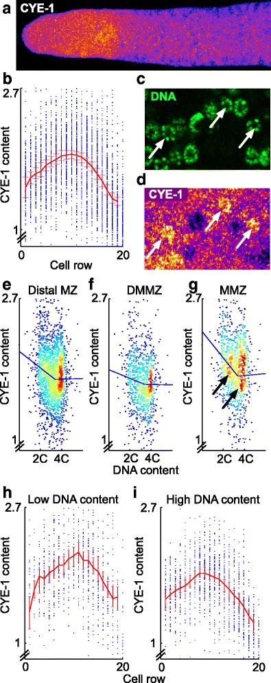
Fig. 5.

Cyclin E levels are graded across the DMMZ and MMZ, and are differentially dependent on cell cycle phase in the DMMZ and MMZ. a Example of CYE-1 staining pattern in a gonadal arm at L4 + 1 day (color-coded using ImageJ’s “Fire” lookup table). CYE-1 levels appear to start low in the distal region, rise, and then fall in the proximal region. b Quantification of nuclear CYE-1 levels using 7508 cells segmented from 30 gonadal arms. Each dot represents a cell; the red line is the average at each cell row, with a 95 % bootstrapped confidence interval. c, d Cells with typical G1 morphology (arrows in c) have higher CYE-1 content than their neighbors (d; arrows point to same G1 cells as in c). e Scatterplot of nuclear CYE-1 content vs. DNA content, showing that cells with lower DNA content – i.e. early in the cell cycle – have moderately higher levels of CYE-1 than cells with higher DNA content. Density colored via “jet” lookup table (red: high density, blue: low density), and piecewise-linear trend line computed as described in “Methods”. f, g Variation of CYE-1 content with cell cycle phase is lesser for cells in the DMMZ (f; virtually flat trend line) than in the MMZ (g; steeper trend line). The difference between DMMZ and MMZ is statistically significant (95 % bootstrapped CI for difference in slopes of first component of trend lines: 0.024–0.38, n = 50,000 replicates). Arrows show two clusters at low and high DNA content. h, i Quantification of nuclear CYE-1 profile as in (a), but considering only cells with low (h) or high (i) DNA content
