
Figure 3.
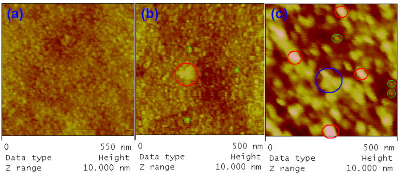
(a) AFM height image of 2k PEG-OH and 3.4k PEG-Mal modified ELISA plate, conducted in air with the tapping mode. (b) AFM height image of dendrimer 4 attached on PEG modified substrate, conducted in air with the tapping mode. (c) A typical AFM height image of antibody-dendrimer conjugate attached on PEG modified ELISA plate. The image was conducted in tapping mode in liquid cell filled with PBS pH 7.4.