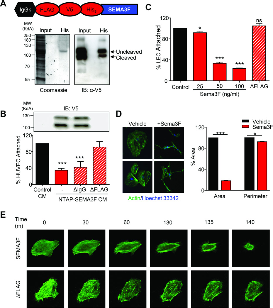
Fig. 2. SEMA3F is a chemorepulsant that negatively impacts the function of lymphatic endothelial cells.
A) Coomassie staining and Western blot of serum-free CM from cells transfected with NTAP-SEMA3F. B) Attachment of HUVEC cells CM from NTAP-SEMA3F cell supernatants alone or after FLAG or IgG immunoprecipitation depletion. C) Attachment of LECs in the presence of increasing amounts of purified SEMA3F or supernatant after FLAG depletion. D) Immunofluorescent staining of actin (green) and nuclei (blue) in LEC treated with 100 ng/ml SEMA3F for 6 hours. Quantification of cell area and perimeter were determined using ImageJ using twenty-five independent fields. Statistical significance was determined using one-way ANOVA, *p<0.05, **p<0.01, ***p<0.001. E) Still images captured during live-cell imaging of LEC transfected with LifeAct GFP and treated with 1 µg/ml SEMA3F or ΔFLAG.

