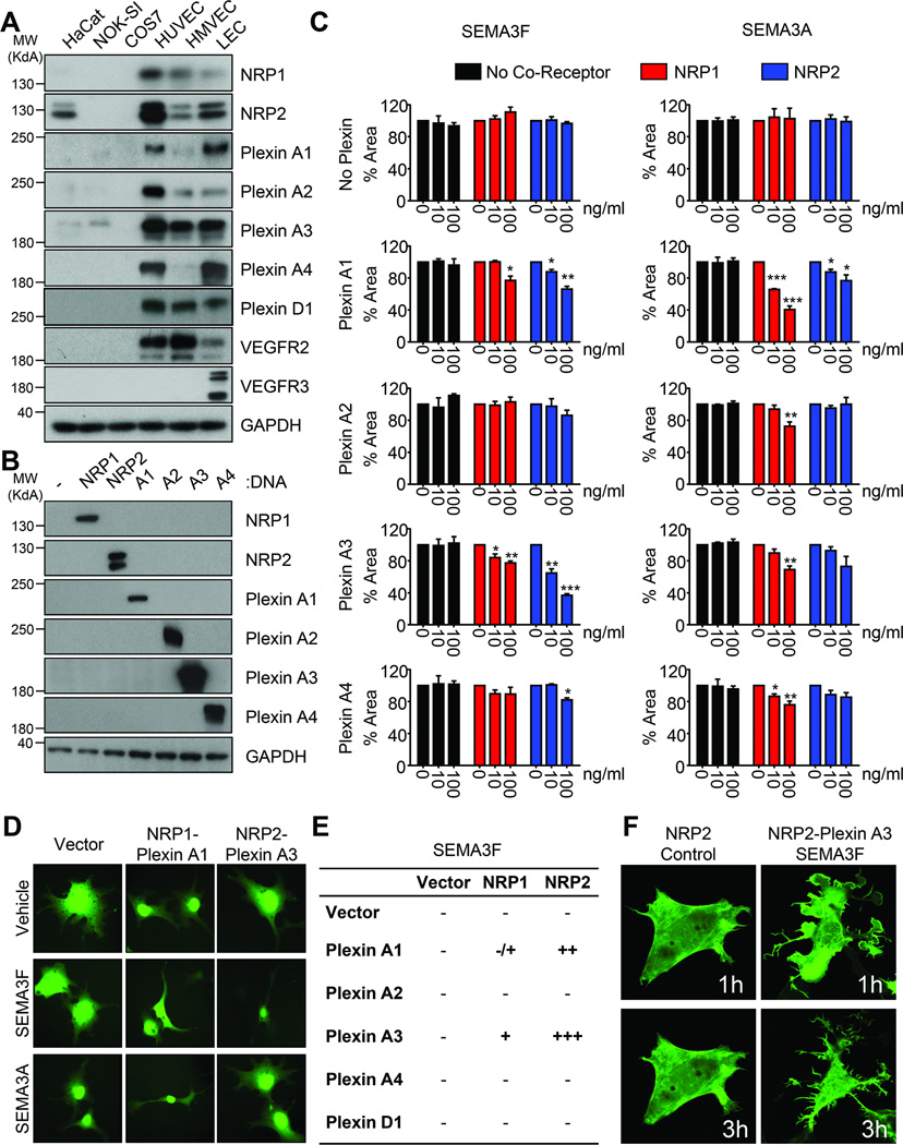
Fig. 4. NRP and Plexin A co-receptors coordinate SEMA3F function.
A) Western blot demonstrating expression of the NRP, Plexin, and VEGFR family members in a panel of immortalized and primary epithelial and endothelial cells. B) Western blot demonstrated specific expression of each NRP and Plexin A family member in COS-7 cells. C) COS-7 cells stably expressing GFP were transfected and treated as indicated. Collapse was quantified using ImageJ relative to vehicle control based on 25 imaging fields each from quadruplicate wells in three independent experiments. Statistical significance was determined using one-way ANOVA, *p<0.05, **p<0.01, ***p<0.001. D) Representative images of cells transfected with different receptor combinations and treated with vehicle, SEMA3F, or SEMA3A. E) Table summarizing the relative contributions of the different receptor combinations tested. F) Confocal images of COS-7 cells transfected with LifeAct-GFP, NRP2, and/or Plexin A3 and treated with SEMA3F or ΔFLAG. Images were taken one hour and three hours after treatment with SEMA3F.

