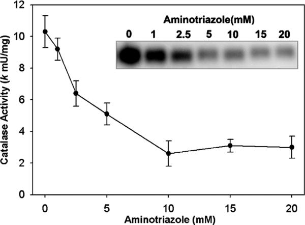
Fig. 1.
Inhibition of intracellular catalase activity by 3-aminotriazole in PC3 cells. PC3 cells were incubated for 2 h with aminotriazole in MEM media with 10% FBS at 37 °C. Cells were rinsed and scrape-harvested into 50 mM phosphate buffer, pH 7.0. Catalase activity was determined by the spectrophotometric catalase assay using 400 μg total cellular protein. Shown is the mean ± SEM of three experiments. Inset: in a complementary method, catalase activity was analyzed by activity gels loaded at 25 μg of PC3 protein per well and stained for catalase activity by the Prussian blue deposition method. Shown is an inverse black/white image of a representative gel.

