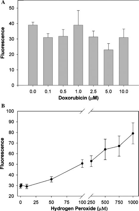
Fig. 5.
(A) Doxorubicin had no effect on carboxy-H2DCFDA fluorescence of human prostate cancer cells. PC3 prostate cancer cells (7.5 × 10⁵) were exposed to increasing concentrations of doxorubicin for 30 min, and 0.25% trypsin and 0.11% EDTA were added during the last 10 min to detach the cells. Carboxy-H2DCFDA was then added for 15 min, fluorescence read on a FACScan flow cytometer (488 nm excitation, 530 nm emission) and corrected for fluorescence contributions by doxorubicin. Shown is geometric mean fluorescence, and values are the mean and SEM of four separate experiments. There was no significant difference by one-way ANOVA (p = 0.3). (B) Carboxy-H2DCFDA fluorescence increases as PC3 cells are exposed to externally applied H2O2. PC3 cells were detached, exposed to carboxy-H2DCFDA for 15 min, and then the desired H2O2 concentrations for 5 min. Carboxy-H2DCFDA fluorescence was measured by flow cytometry. Shown is geometric mean fluorescence, and the data represents the means (±SEM) for three independent determinations. The H2O2 concentrations were significantly different (p < 0.0001) by one-way analysis of variance.

