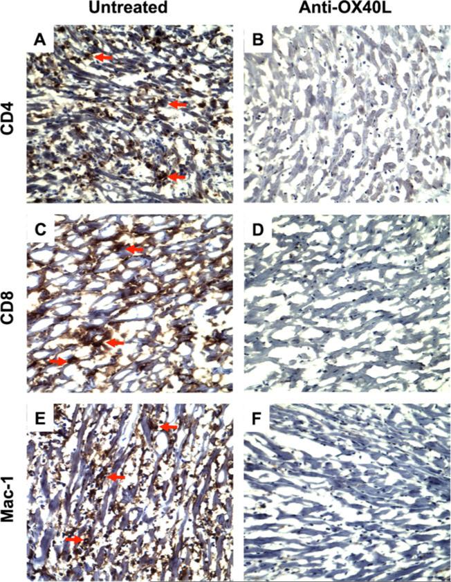
Fig. 4.
Regulation of cellular infiltration (CD4 and CD8 Tcells, and macrophages) in heart allogarfts by anti-OX40L mAb treatment. Cardiac allografts in Rag-1−/− B6 recipients were harvested on POD 100. The CD4, CD8 and Mac-1 expressing cells in the sections of the grafts were localized by immunohistochemistry. Data are presented as a representative immunoperoxidase staining for each of CD4+, CD8+, and Mac-1+ cells. a, c and e A typical microscopic view of CD4+ T cells, CD8+ T cells, or Mac-1+/macrophages in the sections of the grafts from Rag-1−/− recipients harboring Tmem cells (Group 2, untreated, n =8). The red arrows indicated positively stained (brown) cells. b, d and f A typical microscopic view of CD4, CD8, or Mac-1 staining in the sections of the grafts from Rag-1−/− recipients harboring Tmem cells and receiving anti-OX40L mAb treatment (Group 3, n =8). Cytoplasm: blue. Nucleus: dark blue

