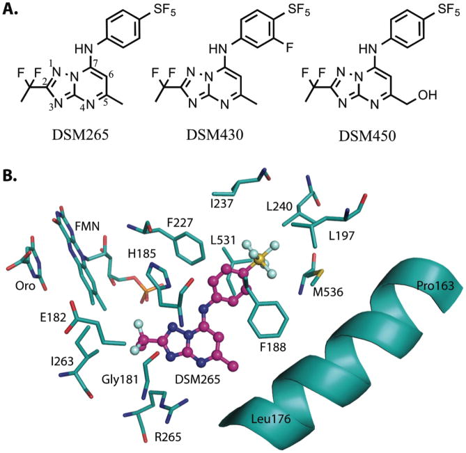
Fig. 1. Chemical and protein bound inhibitor structures.
(A) Chemical structures of DSM265 (415 Da), DSM430 (430 Da) and DSM450 (431 Da). (B) X-ray structure of the inhibitor binding-site of PfDHODH bound to DSM265 (PDB 5BOO). DSM265 (pink); protein, orotate (Oro) and flavin (FMN) (carbons in teal); oxygen (red); nitrogen (blue); sulfur (yellow); phosphate (orange); fluorine (light blue).

